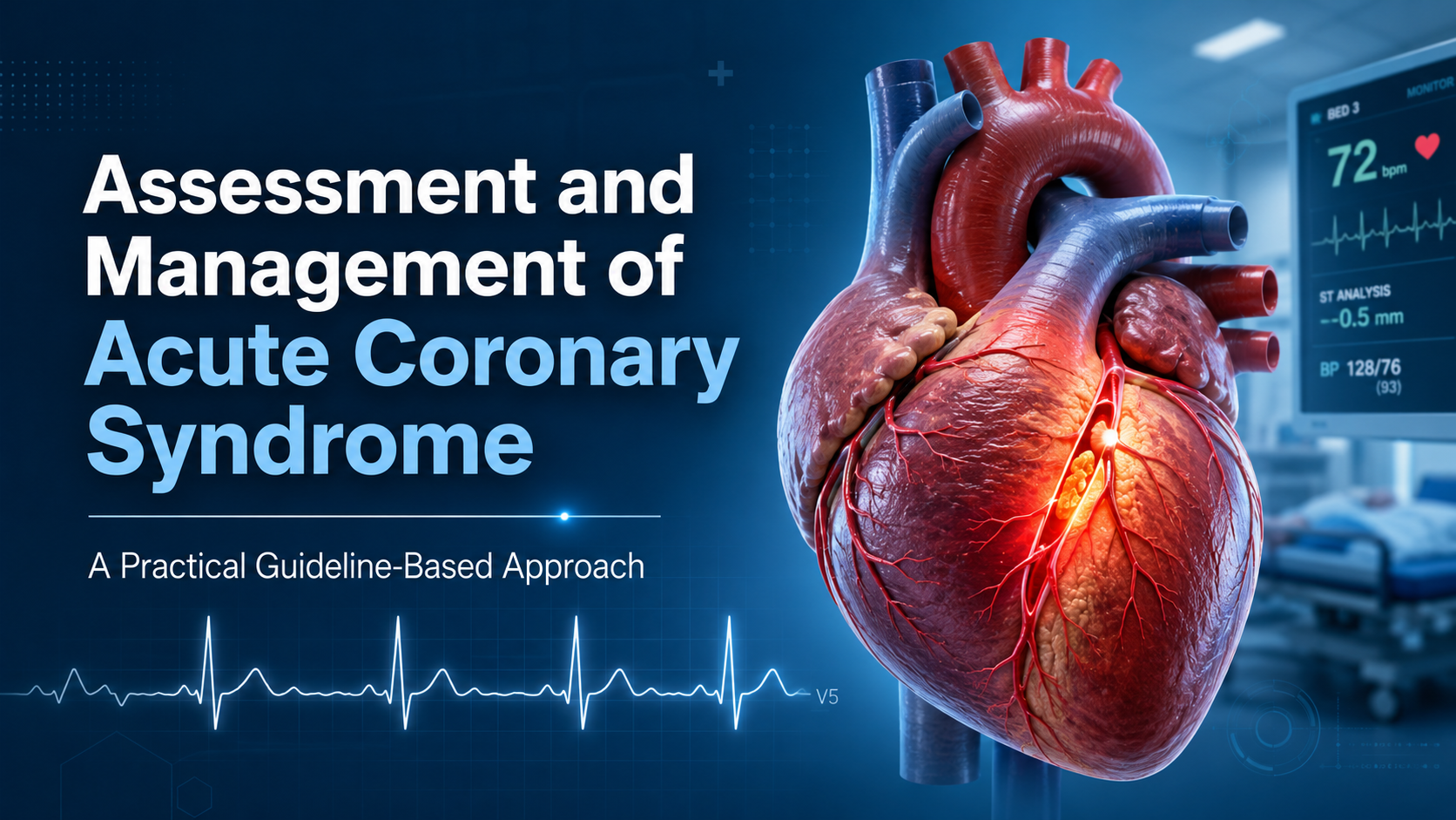
Acute coronary syndrome (ACS) is a time-critical clinical emergency caused by acute myocardial ischaemia, usually due to coronary plaque rupture or erosion with thrombosis. It includes ST-elevation myocardial infarction (STEMI), non-ST-elevation myocardial infarction (NSTEMI), and unstable angina. Modern ACS care is built around one simple principle: identify patients early, restore coronary blood flow when needed, prevent further thrombosis, and begin secondary prevention before discharge. This article summarizes key practical points from the 2025 ACC/AHA/ACEP/NAEMSP/SCAI ACS guideline and the 2023 ESC ACS guideline.
1. Suspect ACS early
ACS should be considered in any patient with acute chest pain, chest tightness, epigastric discomfort, unexplained dyspnoea, syncope, diaphoresis, or haemodynamic instability. Classical central crushing chest pain radiating to the left arm or jaw is important, but ACS can present atypically, especially in older adults, women, people with diabetes, chronic kidney disease, or frailty.
The first clinical task is not to “prove” ACS immediately, but to risk-stratify rapidly and avoid missing a patient who needs urgent reperfusion.
2. Immediate assessment: ABCDE, ECG, monitoring, and vascular access
Initial assessment should include:
- Airway, breathing, circulation, disability, and exposure assessment.
- Vital signs, oxygen saturation, haemodynamic status, and signs of heart failure or shock.
- Focused history: onset, character and duration of pain, associated symptoms, cardiovascular risk factors, prior coronary disease, medications, bleeding risk, anticoagulant use, and contraindications to fibrinolysis.
- Cardiac monitoring and defibrillator availability in unstable patients.
- IV access and urgent blood sampling.
A 12-lead ECG is the key first test. If the initial ECG is nondiagnostic, ACS is not excluded; ECG changes may evolve, and repeat ECGs are recommended when symptoms persist or recur. The ACC/AHA guideline highlights that high-sensitivity cardiac troponin is preferred for myocardial injury assessment, but reperfusion in clear STEMI should not be delayed while waiting for troponin results.
3. ECG-based classification: STEMI or NSTE-ACS
The initial ECG separates patients into two broad pathways.
STEMI or STEMI-equivalent pattern
This includes persistent ST elevation or other ECG patterns suggesting acute coronary occlusion. These patients need immediate reperfusion therapy.
Important STEMI-equivalent patterns include posterior MI, new or presumed new left bundle branch block with ischaemic features, hyperacute T waves, de Winter pattern, and diffuse ST depression with ST elevation in aVR suggesting left main or severe multivessel ischaemia.
NSTE-ACS
This includes NSTEMI and unstable angina. ECG findings may include ST depression, transient ST elevation, T-wave inversion, or even a normal ECG. Diagnosis depends on symptoms, ECG evolution, and serial high-sensitivity troponin testing.
4. Troponin: use high-sensitivity assays and serial testing
High-sensitivity cardiac troponin is the preferred biomarker. A single elevated troponin means myocardial injury, not automatically type 1 MI. The diagnosis of acute MI requires a rise and/or fall in troponin with at least one value above the 99th percentile, together with clinical evidence of acute myocardial ischaemia.
The ESC guideline supports rapid rule-in/rule-out algorithms using 0 h/1 h or 0 h/2 h high-sensitivity troponin pathways in patients with suspected NSTEMI who do not need immediate invasive angiography.
Common causes of troponin elevation other than type 1 MI include sepsis, tachyarrhythmia, heart failure, pulmonary embolism, myocarditis, renal disease, stroke, and severe anaemia. Therefore, troponin should always be interpreted with the clinical picture and ECG.
5. Initial medical treatment
Oxygen
Oxygen should be given to patients with hypoxaemia. The ESC guideline recommends oxygen when SaO₂ is <90%, but routine oxygen is not recommended when oxygen saturation is >90%.
Nitrates
Sublingual or intravenous nitroglycerin can be used for ongoing ischaemic pain, hypertension, or pulmonary oedema, provided the patient is haemodynamically stable. Avoid nitrates in hypotension, suspected right ventricular infarction, severe aortic stenosis, or recent phosphodiesterase-5 inhibitor use. The ACC/AHA guideline gives practical nitrate dosing and cautions, including avoiding nitrates after recent sildenafil, vardenafil, tadalafil, or avanafil use.
Analgesia
Persistent severe pain increases sympathetic drive. Morphine or fentanyl may be considered when pain is resistant to anti-ischaemic therapy, but opioids can delay absorption and effect of oral P2Y12 inhibitors.
6. STEMI management: reperfusion is the priority
For STEMI, the key question is: Can primary PCI be performed quickly?
The ESC guideline recommends reperfusion therapy for all patients with a working diagnosis of STEMI and symptoms of ischaemia of ≤12 hours. Primary PCI is preferred if it can be achieved within the required time window; if PCI cannot be performed within 120 minutes, fibrinolysis is recommended within 12 hours of symptom onset if there are no contraindications.
The ESC pathway emphasises immediate transfer to a PCI-capable centre where feasible. If fibrinolysis is given, the patient should still be transferred to a PCI centre without waiting for signs of reperfusion. Rescue PCI is required if fibrinolysis fails, such as <50% ST-segment resolution at 60–90 minutes, persistent chest pain, worsening ischaemia, or haemodynamic/electrical instability.
The ACC/AHA guideline similarly emphasises regional STEMI systems of care to reduce total ischaemic time and improve survival.
7. NSTE-ACS management: risk determines timing of angiography
NSTE-ACS does not usually require fibrinolysis. Instead, management depends on risk.
Very high-risk NSTE-ACS: immediate invasive strategy
Immediate angiography is recommended when there is:
- Haemodynamic instability or cardiogenic shock.
- Recurrent or refractory chest pain despite treatment.
- Life-threatening arrhythmia or cardiac arrest.
- Acute heart failure due to ongoing ischaemia.
- Mechanical complications.
- Recurrent dynamic ST-segment or T-wave changes, especially intermittent ST elevation.
- High-risk NSTE-ACS: early invasive strategy
High-risk features include confirmed NSTEMI by high-sensitivity troponin algorithm, GRACE score >140, dynamic ST/T changes, or transient ST elevation. These patients should undergo an inpatient invasive strategy and are generally considered for angiography within 24 hours.
Lower-risk NSTE-ACS
Patients without high-risk features may undergo selective invasive assessment after further clinical risk stratification, serial ECGs, troponins, and non-invasive testing where appropriate.
8. Antiplatelet therapy
Aspirin should be started as soon as possible unless contraindicated. Long-term low-dose aspirin, usually 75–100 mg daily, is supported for maintenance therapy.
Dual antiplatelet therapy (DAPT) is central to ACS treatment. The 2025 ACC/AHA guideline recommends DAPT for ACS, with ticagrelor or prasugrel preferred over clopidogrel in patients undergoing PCI, unless contraindicated. The default strategy is DAPT with aspirin plus an oral P2Y12 inhibitor for at least 12 months in patients who are not at high bleeding risk.
The ESC guideline similarly recommends a default DAPT strategy after ACS, usually aspirin plus prasugrel or ticagrelor for 12 months, while allowing shortening, extension, or de-escalation depending on bleeding and ischaemic risk.
In NSTE-ACS, routine pretreatment with a P2Y12 inhibitor before coronary anatomy is known is not recommended when early invasive angiography is planned, but pretreatment may be considered if angiography will be delayed and bleeding risk is not high.
9. Anticoagulation
Parenteral anticoagulation is recommended in ACS to reduce thrombotic complications.
For ACS undergoing PCI, unfractionated heparin remains a standard option. The ACC/AHA guideline recommends UFH for ACS patients undergoing PCI and notes that bivalirudin is a useful alternative in STEMI PCI to reduce mortality and bleeding, while fondaparinux should not be used to support PCI because of catheter thrombosis risk.
The ESC guideline also recommends parenteral anticoagulation in STEMI undergoing primary PCI, with UFH as default and enoxaparin or bivalirudin as alternatives; fondaparinux is not recommended for STEMI primary PCI. For NSTE-ACS, UFH is recommended for immediate or early angiography, while fondaparinux is preferred when early angiography is not anticipated, with UFH added at PCI.
10. Revascularization strategy and procedural considerations
For patients undergoing PCI, radial arterial access is generally preferred because it reduces bleeding and vascular complications. The ACC/AHA guideline highlights radial access and intracoronary imaging for complex lesions as key procedural strategies.
In multivessel coronary artery disease, complete revascularisation is generally recommended or considered, depending on the clinical context. In STEMI with multivessel disease and no cardiogenic shock, complete revascularisation may be performed during the index procedure or staged. However, in ACS with cardiogenic shock, culprit-vessel PCI is prioritised and routine immediate PCI of non-infarct-related arteries is not recommended.
11. Cardiogenic shock and unstable ACS
ACS complicated by cardiogenic shock is a medical emergency. Management includes:
- Immediate recognition: hypotension, cold peripheries, oliguria, altered mentation, pulmonary oedema, raised lactate.
- Early activation of cardiology, intensive care, and catheterisation laboratory teams.
- Urgent revascularisation of the culprit artery.
- Vasopressors/inotropes when needed.
- Echocardiography to assess LV/RV function and mechanical complications.
- Selective use of mechanical circulatory support in experienced centres.
The ACC/AHA guideline notes that microaxial flow pump support may be reasonable in selected patients with AMI-related cardiogenic shock, but complications such as bleeding, limb ischaemia, and renal failure must be considered.
12. In-hospital care
During admission, patients need:
- Continuous rhythm monitoring, especially early after MI.
- Assessment for heart failure, arrhythmias, recurrent ischaemia, bleeding, renal dysfunction, and mechanical complications.
- Echocardiography to assess LV function and complications.
- Anaemia and bleeding risk assessment.
- Early mobilisation when stable.
- Review of diagnosis: type 1 MI, type 2 MI, myocarditis, Takotsubo syndrome, pulmonary embolism, aortic dissection, or MINOCA.
Emergency transthoracic echocardiography is recommended when cardiogenic shock or mechanical complications are suspected. Routine early coronary CT angiography is not recommended in patients with suspected ACS when ACS remains the working diagnosis.
13. Secondary prevention before discharge
Discharge planning should begin early. Every patient should leave hospital with a clear diagnosis, explanation of treatment, medication plan, follow-up plan, and warning symptoms.
Lipid lowering
High-intensity statin therapy is recommended for all ACS patients unless contraindicated. The 2025 ACC/AHA guideline recommends adding non-statin lipid-lowering therapy if LDL-C remains ≥70 mg/dL despite maximally tolerated statin therapy, and considers further intensification for LDL-C 55–69 mg/dL in high-risk patients already on maximally tolerated statin therapy. Ezetimibe may be started concurrently with statin therapy.
Beta-blockers, ACE inhibitors/ARBs, and mineralocorticoid receptor antagonists
Beta-blockers are used particularly in patients with reduced LV systolic function, ongoing ischaemia, arrhythmia, or hypertension, provided there is no acute heart failure, shock, severe bradycardia, or contraindication.
ACE inhibitors or ARBs are important after MI, especially with LV systolic dysfunction, hypertension, diabetes, or chronic kidney disease. Mineralocorticoid receptor antagonists are considered in selected patients with LV dysfunction and heart failure or diabetes, provided renal function and potassium allow.
Diabetes and cardiometabolic risk
In patients with diabetes and ACS, glucose-lowering therapy should be individualised. SGLT2 inhibitors and GLP-1 receptor agonists with cardiovascular benefit are important considerations in long-term care, especially in patients with type 2 diabetes, chronic kidney disease, heart failure, or established atherosclerotic cardiovascular disease.
Cardiac rehabilitation
Cardiac rehabilitation is a core component of ACS care. The ACC/AHA guideline recommends referral to cardiac rehabilitation, including home-based programmes where in-person attendance is not possible. It also recommends reassessing fasting lipids 4–8 weeks after starting or adjusting lipid-lowering therapy.
14. Practical ACS pathway
A simple bedside approach is:
- Recognise possible ACS from symptoms, risk factors, and clinical instability.
- Obtain ECG immediately and repeat if nondiagnostic or symptoms persist.
- Classify as STEMI/STEMI-equivalent or NSTE-ACS.
- For STEMI: activate primary PCI pathway; use fibrinolysis if PCI cannot be achieved within the recommended time and there are no contraindications.
- For NSTE-ACS: use serial high-sensitivity troponin, ECG changes, haemodynamic status, and GRACE score to decide immediate, early, inpatient, or selective invasive strategy.
- Start antithrombotic therapy: aspirin, appropriate P2Y12 inhibitor strategy, and parenteral anticoagulation.
- Treat symptoms safely: oxygen only if hypoxaemic, nitrates if appropriate, analgesia when needed.
- Assess complications: shock, heart failure, arrhythmias, mechanical complications, bleeding.
- Plan secondary prevention: DAPT strategy, statin ± ezetimibe/non-statin therapy, BP and diabetes control, smoking cessation, cardiac rehabilitation, follow-up.
Key messages
ACS management is a race against time, but it is also a process of careful risk stratification. STEMI requires rapid reperfusion. NSTE-ACS requires early identification of high-risk patients who benefit from invasive management. Antiplatelet therapy, anticoagulation, lipid lowering, cardiac rehabilitation, and patient education are not “discharge extras”; they are essential treatments that reduce recurrent MI, stroke, and death.
References
- Rao SV, O’Donoghue ML, Ruel M, Rab T, Tamis-Holland JE, Alexander JH, et al. 2025 ACC/AHA/ACEP/NAEMSP/SCAI Guideline for the Management of Patients With Acute Coronary Syndromes: A Report of the American College of Cardiology/American Heart Association Joint Committee on Clinical Practice Guidelines. Circulation. 2025;151:e771–e862. doi:10.1161/CIR.0000000000001309.
- Byrne RA, Rossello X, Coughlan JJ, Barbato E, Berry C, Chieffo A, et al. 2023 ESC Guidelines for the management of acute coronary syndromes. European Heart Journal. 2023;44:3720–3826. doi:10.1093/eurheartj/ehad191.